The Sodium Fluorescein technique:
Light sources and Filtration
The xenon tubed electronic flashgun is again, the preferred light source. It has a good output of radiation at the 450nm frequency required to stimulate fluorescein and has the merit of being of very short duration - critical for in vivo medical applications. Often a hand lamp with high output in the blue region is most useful for examining the fluorescing area prior to photography - although it has to be said that with fluorescein in general - 'if you can't see it you won't be able to photograph it' is an apposite phrase. The fluorescence should be visible to the naked eye - the clarity of visualisation will then be enhanced enormously by using the appropriate barrier filter and extinguishing all 'ambient' lighting. The spectral details for electronic flash and details of continuous sources of blue/ultraviolet radiation can be found in the section on 'Sources of ultraviolet radiation' in Reflected Ultraviolet Photography.
The excitation filter (deep blue) is usually a Wratten 47 or 47A and has to be fitted into a light-tight mount over the front of the light source. The spectral response curves for the Wratten 47 and 47A filters are shown in Figures 38 and 39. The 47A has been especially formulated to stimulate sodium fluorescein.
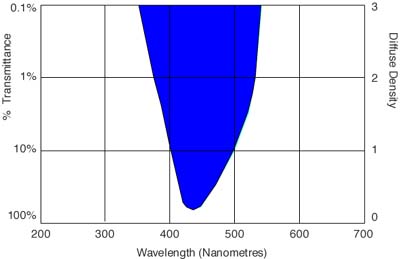
Figure 38 (above). The spectral transmission curve for the Wratten 47 deep blue filter.
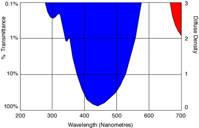
Figure 39 (above). The spectral transmission curve for the Wratten 47A filter especially formulated for stimulation of sodium fluorescein dye.
Fundus cameras, however, often use high quality interference filters, eg., the Spectrotech SE40P, with a narrower transmission centred on the 450nm frequency. Some workers have claimed superior results in all their fluorescein patient photography by using these high quality, narrow-cut interference filters, eg., Erol (1981). Others have claimed the results from the 47A and the Spectrotech SE40P to be very similar (Lanzafame et al., 1982).
The standard barrier filter is a deep yellow or orange - a Wratten 12 or 15 - which eliminates all the reflected blue light. The spectral transmission curves for these are shown in Figures 40 and 41 respectively. Often a Wratten 2B or 2E ultraviolet absorbing filter is added to this barrier filter. Figure 42 show the combinations of the Wratten 47A with the Wratten 12 and Figure 43 show the combination of the Wratten 47A with the Wratten 15 - these are the most popular choices for excitation and barrier filters. Hodge (1978) discussed extensively all the possible filter combinations and their respective windows of transmission.
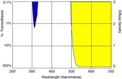
Figure 40 (above). The spectral transmission curve for the Wratten 12 yellow barrier filter.
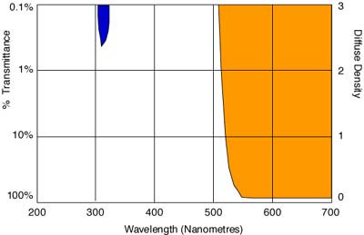
Figure 41 (above). The spectral transmission curve for the Wratten 15 orange barrier filter.
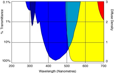
Figure 42 (above). Shows the combination of the Wratten 47A excitation filter with the Wratten 12 barrier filter.
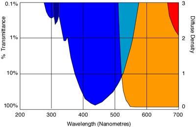
Figure 43 (above). Shows the combination of the Wratten 47A excitation filter with the Wratten 15 barrier filter.
References
- Erol, 0. and Spira, M., 1981, "An improved method for fluorescein dye photography," Plast. Reconstr. Surg. 68 (1):120-121.
- Hodge, C., 1978, "Fluorescein angiography of the brain - the photographic procedure," J. Biol. Photogr. 46(2):67- 79.
- Lanzafame, R. et al. , 1982, "Fluorescein photography: A method for documentation of tissue perfusion using instant photography," Current Surgery 39:310- 313.
|
