about | articles | authors | contact | links
about | articles | authors | contact | links |
![]() Home > Articles > Reflected Ultraviolet Photography > Sources of ultraviolet radiation
Home > Articles > Reflected Ultraviolet Photography > Sources of ultraviolet radiation
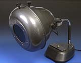
REFLECTED ULTRAVIOLET PHOTOGRAPHYAuthors: Prof. Robin Williams and Gigi Williams Sources of ultraviolet radiationThere are many sources of ultraviolet radiation; indeed sunlight itself emits 10% of its energy in the ultraviolet region (Figure10). Tungsten lamps are very poor sources of ultraviolet (Figure 11) and should be avoided. In practice, ultraviolet sources can be divided into two types - continuous and flash. Continuous sources are more suited to chromatography, immunoelectrophoretic studies, fluorescence and document examination, while flash is obviously more suited to patient photography. Gas discharge lamps, particularly the mercury vapour discharge lamp, are the most popular form of continuous ultraviolet source. The proportion of ultraviolet emitted by a mercury vapour lamp varies considerably with current density, but the operating pressure of the lamp mainly governs the spectral quality. Low-pressure mercury vapour lamps emit about 90% of their output as a line spectra at 254nm (Figure 12). High-pressure lamps, however, have a peak output at 365nm and secondary peaks at 334nm and 313nm (Figure 13). The high-pressure mercury vapour lamp fitted with a Wood's filter (Figure 14), is the standard instrument in dermatology for examining the skin under ultraviolet-this filter virtually restricts output of the lamp to a line at 365nm.
Figure 10 (above). The spectral emission for averaged daylight shows a reasonable amount of ultraviolet content (Note this may vary enormously depending on the weather, time of day/year and the latitude).
Figure 11 (above). The spectral emission for Tungsten illumination demonstrates that whilst it is relatively rich in infrared it is a very poor source of ultraviolet radiation.
Figure 12 (above). The spectral emission of a low pressure mercury vapour lamp is a line spectra with about 90% of its output at 254nm.
Figure 13 (above). High-pressure lamps, however, have a peak output at 365nm and secondary peaks at 334nm and 313nm
Mercury vapour lamps are supplied by several companies; in the USA these are:
The domestic fluorescent tube is a very poor source of ultraviolet but special tubes are available with ultraviolet emitting phosphors - the so-called "black light" tubes. They are inexpensive, efficient sources of ultraviolet and do provide very even illumination that is useful for large subjects. They are, however, quite inefficient when compared with the output of either mercury vapour or xenon flash tubes. Some manufacturers incorporate small ultraviolet fluorescent tubes into battery driven hand lamps - these are very useful as examination lamps prior to fluorescence photography with a more efficient source (covered in more detail in fluorescence photography). Open arcs provide substantial emission of ultraviolet and are still used as primary sources in some process work. The xenon arc lamp is a particularly good continuous source of ultraviolet. It has a fairly flat, but high spectral output from 300nm to 1100nm and is therefore suitable as a continuous source for both ultraviolet and infrared photography. Figure 15 shows the spectral emission curve for the xenon arc lamp.
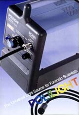
Figure 15 (above). The xenon arc lamp is a particularly good continuous source of ultraviolet. It has a fairly flat, but high spectral output from 300nm to 800nm with a peak of activity between 800nm and 1100nm and is therefore suitable as a continuous source for both ultraviolet and infrared photography. Several manufacturers now supply xenon arc lamps especially made for invisible radiation work. Examples would be the Polilight, Lumilite or Omniprint. These all have a series of stepped interference filters, which are then tunable by tilting their angle to the beam to give a continuously variable output from 300 to 1100nm. An LED display shows the frequency of the output accurate to +/- 20nm. Visible and ultraviolet radiation are delivered to the point of use by an efficient liquid light guide with quartz and silica optics on the end and infrared via conventional fiber-optic light guide (Figure 16).
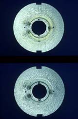
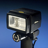
The xenon flash discharge tube has a very high output between 300 and 400nm and is the most useful source of ultraviolet to the biomedical photographer. Figure 17 shows the spectral output of the standard xenon flash tube. Electronic flash manufacturers have been well aware of the high output of ultraviolet radiation and the deleterious effect this has on colour photography (blue colour casts in shadow areas). In many instances they have coated their flash tubes with a gold coating to absorb the ultraviolet or have fitted ultraviolet absorbing screens to the front of the flashgun (Figure 18). The effect of such filtration is quite variable; some coatings seem very efficient and markedly reduce ultraviolet output, while others have very little effect. Ultraviolet absorbing filters fade with exposure and thus become ineffective over time and this may explain why older or well-used flash tubes suffer less from the absorption problem. When new, however, such coatings do absorb ultraviolet efficiently, and so it is helpful to obtain uncoated or unfiltered tubes. Several manufacturers supply them for studio flashes (Bowens, Courtney and Elinchrom). Nikon used to supply a portable flash with an uncoated tube - the SB-140 (Figure 19). The Nikon unit came complete with filters for both ultraviolet and infrared work and had a useful exposure guide - in many respects the ideal source for invisible radiation patient photography, especially on location. Figures 20 to 23 show the spectral emission curves for this source with its various filters. Unfortunately Nikon have now withdrawn this unit from their product range - like so many products suited to specialised imaging from different manufacturers. The authors are fortunate enough to own two of these SB-140s and can vouch for their usefulness for invisible radiation photography; so don't hesitate to acquire one if you happen to come across one second hand or 'left over' in a camera store.
Figure 17 (above). The spectral output of the standard xenon flash tube with a very high output between 300 and 400nm this is the most useful source of ultraviolet to the biomedical photographer.
Figure 20 (above). The spectral emission curve for the unfiltered Nikon SB-140 xenon flash tube.
Figure 21 (above). The spectral emission curve for the Nikon SB-140 xenon flash tube fitted with its UV transmission filter.
Figure 22 (above). The spectral emission curve for the Nikon SB-140 xenon flash tube fitted with its infrared transmission filter.
Figure 23 (above). The spectral emission curve for the Nikon SB-140 xenon flash tube fitted with a filter designed for 'normal' visible light photography. Great care needs to be taken when working with continuous ultraviolet sources or a severe burn to the patient, or a keratitis to the photographer, may occur. As can be seen from Figure 4 the peak of erythemal activity occurs at about 310nm. Conjunctivitis and skin erythema only appear 4 to 5 hours after exposure to ultraviolet radiation, and there is no heat involved to warn the photographer of an impending burn. In addition, the retina only starts "seeing" above 400nm, so there is a real potential danger to both photographer and patient. The low-pressure mercury vapour lamp has found renewed popularity in an unfiltered form for some electrophoretic investigations using radiation at 254nm. Special care needs to be taken with these and with the newer short-wave ultraviolet fluorescent tubes - goggles and skin protection must be worn. Electronic flash only emits a very brief burst of radiation and is completely safe - it is therefore a much more practical source for patient photography. The medical photographer however should be aware that patients suffering from xeroderma pigmentosum can be seriously affected by even moderate exposure to normal electronic flash (Menezes, 1996). References
|
| © 2002 Prof. Robin Williams and Gigi Williams - Disclaimer URL: http://www.medicalphotography.com.au/Article_01/ Last modified: 3 May 2002 |