The Sodium Fluorescein technique:
Introduction
While many substances fluoresce naturally, it is also helpful to be able to "label" tissues and chemicals with dye markers, or fluorochromes, and then examine the secondary fluorescence that results. Many of these fluorochromes fluoresce under stimulation by ultraviolet radiation, but some, particularly those derived from fluorescein, fluoresce much better under blue-light stimulation.
Probably the most commonly used of these dyes in the biological world is sodium fluorescein. Sodium fluorescein is a protein dye, so some caution is required in its use within the living body. This dye is either injected into the bloodstream of the patient in concentrated form from standard vials (Figure 34) or administered externally in some fashion - typically on small strips which are moistened with sterile water (Figure 35). Although the dye will be passed out in the urine after its administration, the body builds up an immune reaction to successive doses of the dye. The immune response is cumulative - so patients should not receive more than the minimum number of investigations with sodium fluorescein. The dye may eventually cause severe anaphylactic shock and even cardiac arrest in some patients. Sodium fluorescein almost always induces a feeling of nausea. Medically qualified help must always be available, and resuscitation facilities at hand when undertaking this kind of photography. Patients must also be warned that their urine will be bright fluorescent yellow for some hours after administration! (Figure 36).
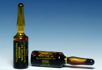
Figure 34 (above). Vials of sodium fluorescein the protein dye fluorochrome used widely in medicine, especially ophthalmology.
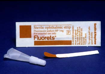
Figure 35 (above). Sodium fluorescein 'Fluorets' - used for external single application such as identification of corneal abrasion. The Fluoret is activated by a few drops of normal saline.

Figure 36 (above). A cartoon illustrating the unfortunate effect of an intravenous Sodium Fluorescein injection - the patient will urinate bright yellow fluorescent urine for several hours after administration.
Many workers have erroneously used ultraviolet radiation to stimulate fluorescein and then subsequently discovered the vastly superior results obtained by using the correct stimulating frequency, eg., Grossman (1981). Sodium fluorescein has its peak absorption at 450nm and produces a yellow/green emission when stimulated by light in the blue region.
There are many other fluorochromes derived from fluorescein, particularly the isothyocyantes, which are often used in tumour biology and to localise antigens and antibodies, eg., fluorescein isothyocyanate or FITC, and Acriflavine S. These are mostly used at cellular level, and so involve the use of the microscope - but the fluorescence technique is essentially the same. Loveland (1981) gives a truly excellent description of fluorescence microscopic technique.
The technique for photographing fluorescein is basically similar to ultraviolet fluorescence; but here the subject has to be illuminated with light in the region 420 to 480nm. The yellow/green fluorescence at about 530 to 540nm is then recorded onto colour film, the blue light being absorbed by a deep /orange barrier filter over the lens. Figure 37 shows the basic technique used. It is critical that one works in a darkened room - surgical lights in the operating theatre, for example, have to be extinguished for the period of visualisation and recording.
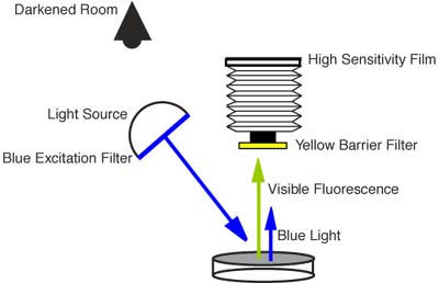
Figure 37 (above). shows the basic sodium fluorescein photographic technique. The subject is illuminated with blue light at 450nm and then recorded onto high speed film through a yellow or orange barrier filter. It is critical that one works in a darkened room.
References
- Grossman, J., 1981, "A simple technique for fluorescein photography," Plast. Reconstr. Burg. 67 (2):257-258.
- Loveland, R., 1981, "Photomicrography -a comprehensive treatise in two volumes" Krieger Publishing Co. Malabar FL. USA in Association with John Wiley.
|
