about | articles | authors | contact | links
about | articles | authors | contact | links |
![]() Home > Articles > Fluorescence Photography > Introduction
Home > Articles > Fluorescence Photography > Introduction
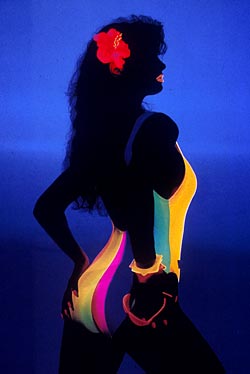
FLUORESCENCE PHOTOGRAPHYAuthors: Prof. Robin Williams and Gigi Williams IntroductionIn order to understand this photographic technique it is important to first describe what fluorescence is. Fluorescence is the stimulation and emission of radiation from a subject by the impact of higher energy radiation upon it. Fluorescence, unlike phosphorescence, ceases almost immediately when excitation is removed. (Luminescence is a general term for the emission of radiation that incorporates both fluorescence and phosphorescence, as well as other electro-chemical phenomena like bioluminescence). Many naturally occurring substances fluoresce, including rocks and minerals, fungi, bacteria and most body tissues; this is termed primary fluorescence or autofluorescence (Jagger, 1967). Alternatively, fluorescent marker dyes, or fluorochromes, can be introduced into a subject when the resulting fluorescence is termed secondary. Marker dyes are frequently used in medicine and forensic science. Dake & DeMent (1942) discussed a whole range of fluorescence chemicals and their applications. Fluorescent materials are widely used: most detergents and washing powders contain optical brighteners, as do many papers and graphic arts products. Plastics and paints often contain fluorescent dyes when used for high visibility applications - such as safety equipment and road signs - and many fashion fabrics and accessories are made of fluorescing materials (Figure 1).
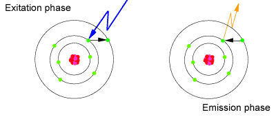
Sir George Stokes published the first significant paper on fluorescence in 1852. Stokes invented the phrase 'fluorescence' after the mineral fluorospar; derived from the Latin fluo (to flow) and spar (rock). He described the theoretical basis for fluorescence. There is a vast wealth of literature available on the fluorescent properties of various materials. A recent comprehensive literature search by the authors revealed some 15,000 titles. Passwater (1967) has published a very helpful guide to this body of knowledge. Stokes's Law states that the incident radiation must always be at a higher energy level (shorter wavelength) than the radiation emitted by any absorber of that radiation (Radley & Grant, 1959). High-energy radiation impinging on the subject causes the electrons of the subject's atoms to be temporarily raised to higher energy orbits. The electrons then naturally slip back to their normal orbit releasing energy as they do so, often in the form of light (Figure 2). The light emitted is always of longer wavelength (lower energy) than the light causing the stimulation because some energy is inevitably used up in the process of getting out to the higher orbit. Thus, subjects irradiated with ultraviolet may release, for example, green, yellow or pink light and subjects irradiated with visible light may emit infrared fluorescence. Theoretically any wavelength of "light" may be used as the stimulating source but in practice it is usually the higher energy sources, ie. the shorter wavelengths, that are used photographically. Guilbault (1973) describes fully the theory of fluorescence.
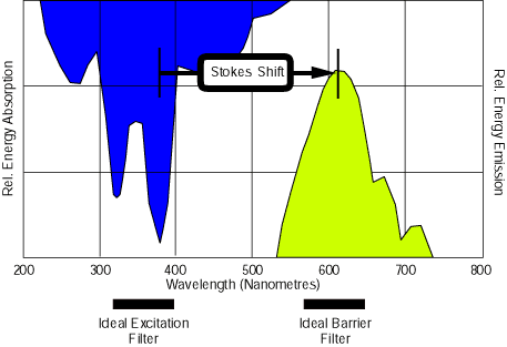
Many biological subjects exhibit the fluorescence phenomenon and have characteristic absorption spectra. By using a spectrophotometer it is possible to record the relative absorption of any substance across a range of wavelengths. Parker (1988) describes the modern range of spectrometers and their use. From such a graph the maximum absorption frequency (or wavelength) can be determined. The aim in fluorescence photography is to stimulate the subject with radiation that corresponds to this maximum absorption frequency - the ideal excitation filter would therefore correspond to the curve for the maximum absorption. This will then often induce some kind of fluorescence at a longer wavelength, and ideally the barrier filter curve should be matched exactly to the peak fluorescence wavelength (Figure 3). In practice this perfect technique would require the use of a spectrophotometer to determine the absorption spectra of the subject and a light source that was "tunable" across a wide range of wavelengths with a comprehensive set of narrow cut barrier filters. These instruments and filters exist, but are costly and usually only found in advanced optical laboratories. In practice, therefore, fluorescence photography is usually limited to one of three techniques: ultraviolet induced emission of natural autofluorescence or of introduced marker dyes (fluorochromes), blue stimulation of dyes such as sodium fluorescein and fluorescein isothyocyanate, and cyan induced of natural autofluorescence which is then emitted in the infrared spectrum.
References
|
| © 2002 Prof. Robin Williams and Gigi Williams - Disclaimer URL: http://www.medicalphotography.com.au/Article_02/ Last modified: 3 May 2002 |