about | articles | authors | contact | links
about | articles | authors | contact | links |
![]() Home > Articles > Pioneers of invisible radiation photography > Charles Hodge
Home > Articles > Pioneers of invisible radiation photography > Charles Hodge
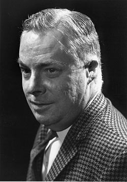
PIONEERS OF INVISIBLE RADIATION PHOTOGRAPHYAuthors: Prof. Robin Williams and Gigi Williams Charles Hodge (1924 - 2001)Charles Hodge (Figure 32) started his career at the Montreal Neurological Institute in Canada in 1945 where he remained as its head of the department of Neurophotography for nearly 50 years. His achievements are many, amongst which he was the first Canadian to be elected a Fellow of the Biological Photographic Association (1958). He was given the first William Gordon Award (1969) for outstanding biophotography in Canada. In 1970 he was winner of the combined Royal Colleges Medal awarded by the Royal College of Physicians, Surgeons, Obstetricians and Gynaecolologists and the Royal Photographic Society of Great Britain and a Louis Schmidt Laureate (1975), the highest honour of the BPA. He became Honorary Fellow of the Royal Photographic Society of Great Britain (1984) and honorary Fellow of the Institute of Medical Illustrators of Great Britain (1990). In 1992 he was appointed to the Order of Canada. Sadly 'Charlie' died in 2001.
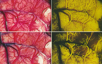
A significant use of fluorescein photography was reported in the Canadian Medical Journal in 1967 by the team Feindel, Yamamato and Hodge - this built on two earlier publications in 1965 by Feindel. The paper described how through a wide craniotomy, fluorescein photographs were taken of the blood supply to the brain. The technique clearly allowed differentiation between arterial and venous vessels and the blood supply to tumours (Figure 33). There was a brief description of the photographic technique based on conventional 1% sodium fluorescein using Tri-X pan with Wratten 47 and 58 filters - this was the first recognition that the optimum excitation of fluorescein was not in the ultraviolet region. In this early work the authors advocated using Ektachrome daylight film rated at 1500ASA. Pictures were taken every 0.4 of a second using a Nikon motor drive camera.
Figure 33 (above). Fluorescein photographs showing differentiation between arterial and venous vessels and the blood supply to tumors in the brain. In 1978, Charles Hodge wrote what is considered to be the definitive work on fluorescein photographic procedures in the Journal of the Biological Photographic Association. His paper was entitled 'fluorescein angiography of the brain - the photographic procedure'. It reported fully the photographic experiments, which had been partially described, by Feindel, Yamamoto and Hodge in 1967. This was a truly excellent paper and examined all of the factors, which affect the photographic result in fluorescein photography. He was the first to note that the concentration of the fluorescein was particularly important and showed that too much, or too little, fluorescein resulted in no significant fluorescence. He also demonstrated very effectively by using a prism that fluorescein had its maximum excitation in the blue region, thus explaining why many previous researchers imagining fluorescein to be an ultraviolet fluorescence technique had achieved poor results, or at least results with very long exposures. Hodge went on to examine all of the possible filter combinations and showed the curves as well as photographic results. At the end of the paper he presented an excellent summary diagram of the possible combinations of excitation and barrier filter. His favourite combination was the use of the Wratten 47A in combination with the 21 barrier filter. He also used a Wratten 2B ultraviolet absorbing filter with the number 21. In addition he recorded details of push processing for Ektachrome 200 film and described the addition of sodium hydroxide to the colour developer to improve image quality. The paper stands out as the most useful and practical description of fluorescein photography and most of the researchers publishing in the medical literature would have done better if they had read Hodge's work. References:
|
| © 2002 Prof. Robin Williams and Gigi Williams - Disclaimer URL: http://www.medicalphotography.com.au/Article_04/ Last modified: 3 May 2002 |