about | articles | authors | contact | links
about | articles | authors | contact | links |
![]() Home > Articles > Pioneers of invisible radiation photography > Professor Ralph Marshall
Home > Articles > Pioneers of invisible radiation photography > Professor Ralph Marshall
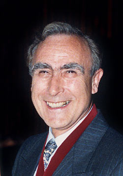
PIONEERS OF INVISIBLE RADIATION PHOTOGRAPHYAuthors: Prof. Robin Williams and Gigi Williams Professor Ralph MarshallRalph Marshall (Figure 30) trained as a medical photographer at the Royal Postgraduate Medical School of the University of London. In 1952 he moved to Wales where he set up a medical illustration service for the Welsh National School of Medicine and the United Cardiff Hospitals. His work as a teacher and researcher was recognized in 1986 when he was appointed professor of Medical Illustration at the University of Wales.
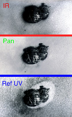
Marshall has been one of the key workers in the field of invisible radiation photography for many years. He has an extensive and varied publication list which began with a joint publication in 1963 with Aldis in which they were able to demonstrate that deep seated malignant melanomas were absorbing infrared radiation. In 1976 Marshall continued to study the effects of invisible radiation on lesions of the skin. Preliminary findings from this research were presented in a paper where he described a comprehensive study of pigmented lesions in both ultraviolet and infrared radiation. In this publication he stated that the infrared photographs tended to separate out the different kinds of lesion, melanoma showing a relatively high degree of correlation with low infrared reflectance. Marshall went on with his studies as part of an academic program of work that led to a doctorate in 1977. In this Doctoral thesis he showed the method to be useful for numerous applications. In his study of specimens for example, he showed that heart muscle was highly reflective in the infrared record whilst the haemorrhagic areas on the lobes of the lung absorbed infrared quite strongly. The small and large intestines were differentiated markedly in the infrared record, while infarcts were highly reflective in the placenta. The infrared record of the neonatal spine demonstrated that underlying detail could be seen, thereby showing details of the nerve roots and the spinal cord not ordinarily visible. However Marshall found that with the reflected ultraviolet technique there was no striking difference to panchromatic photography apart from enhanced surface detail for specimens of the intestine and that there was no advantage for the reflected ultraviolet technique for specimens of the neonatal spine. In his thesis he particularly concentrated on malignant melanoma; but also showed fungating carcinoma of the breast where ultraviolet photography appeared to offer little advantage over panchromatic photography. He demonstrated that ultraviolet could sometimes reveal unexpected changes in skin pigment, which could point to a precancerous lesion, as in Sutton's halo naevus (Figure 31).
He showed that the reflected ultraviolet technique was useful for delineating a hot water bottle burn of the abdomen and for showing comedones but was not useful for showing petechie. This same technique was found to be useful in showing contrast in pityriasis versicolor, but not for xanthelasma palpebrarum. The ultraviolet technique erased tattoos and was not useful for 'miners stripes.' Whereas infrared demonstrated xanthelasma, tattoos and miners stripes very clearly. In 1978 Marshall published a paper on uneven development effects with infrared sheet film and described an agitation technique for removing this effect. Another publication on infrared photography followed a year later in 1979 when he described how the penetrating effects of infrared radiation were useful in revealing neural tube defects. He also published a paper that same year on the usefulness of infrared photography in recording various types of specimen. Further work from the PhD thesis was then presented in 1980 in which he evaluated the usefulness of the photographic photometry as a diagnostic test for malignant melanoma. In this he concluded that a measurement of the difference in absorption of the lesions to infrared and ultraviolet radiation was a more sensitive test than observation by experienced clinicians. In 1981 Marshall built on his original work on pigmented lesions of the skin and its possible clinical significance in malignancy by reporting a large-scale study of many lesions using reflected ultraviolet photography. He concluded that this technique was especially good at recording the depigmentation associated with premalignant lesions. Another paper in the Journal of Audio Visual Media in Medicine in 1981 concerned a further study on the use of infrared and ultraviolet reflectance measurements in the diagnosis of pigmented lesions of the skin. His thesis and subsequent paper in 1980 had indicated that invisible radiation photography was a useful diagnostic test for determining malignant melanoma - this was of such significance that further study was undertaken of 63 pigmented lesions to confirm the value of the technique. In this study his results demonstrated that the reflectance measurements made by photographic photometry may be a less sensitive objective test than observation by an experienced clinician. In his discussion of the apparent disparity between this and earlier studies, Marshall concluded that there was very little difference in the sensitivity of the photographic photometry in both studies, but in this latter study the presence of an experienced diagnostician on the clinical group meant they performed rather better. At the end of this paper Marshall indicated that the level of expertise required for this kind of analysis was considerable and that what was really needed was a simple real-time electronic system for doing the same thing. It was only one year later in 1982 that Marshall published a paper on a television method for measuring infrared and ultraviolet reflectancies of the pigmented lesions. This paper was based on having three television cameras with appropriate tubes and filters feeding out to an oscilloscope with a line selector. A comparison of the density across a lesion was compared with the density across a step wedge. This enabled very accurate real-time measurement of the densities across the pigmented lesions. By 1982 Marshall published his first work on ultraviolet fluorescence photometry. In this study he was studying the rate of skin turnover in the Dansyl chloride test by actually recording the density of the fluorescence of the marker compound. This was shown to be significantly better than a visual comparison with a density chart which had been the previously accepted technique. References:
|
| © 2002 Prof. Robin Williams and Gigi Williams - Disclaimer URL: http://www.medicalphotography.com.au/Article_04/ Last modified: 3 May 2002 |