about | articles | authors | contact | links
about | articles | authors | contact | links |
![]() Home > Articles > Pioneers of invisible radiation photography > Ray Ruddick
Home > Articles > Pioneers of invisible radiation photography > Ray Ruddick
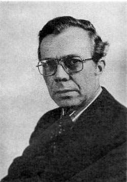
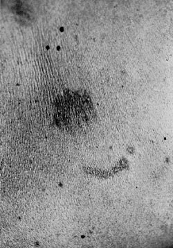
PIONEERS OF INVISIBLE RADIATION PHOTOGRAPHYAuthors: Prof. Robin Williams and Gigi Williams Ray RuddickIn 1950 Ray Ruddick (Figure 34) became Photographer in charge at "The London" (Hospital) where he was he was to remain for his entire working life. Over a period of fifteen years Ray made a particular study of the recording of "hidden lesions", where ultra-violet reflection techniques have enabled records to be made many weeks after all visible signs of trauma have disappeared. In 1973 together with Cameron and Grant, Ruddick published a paper that emphasized the value of reflected ultraviolet photography in forensic medicine with an illustrated example of some old hammer blows to the head. They recommended non-colour sensitive process film for 'new' injuries and true reflected ultraviolet records for old injuries. His 1974 paper published in Medical and Biological Illustration entitled 'a technique for recording bite marks for forensic studies' is widely quoted in forensic circles. In this paper Ruddick described a conventional reflected ultraviolet technique using a Wratten 18A filter and Ilford FP4 and illustrated the article with an example of bite marks (Figure 35). He stated that new injuries were best recorded using a blue sensitive film with no other filtration, and went on to say that there was a period after the initial skin lesion had faded away when it was not possible to record lesions by any method. He then noted that anything between 3 and 6 months after injury the reflective ultraviolet technique was capable of successfully recording old wounds. He explained the phenomenon as being due to the absorption of melanin by ultraviolet and the notion that 'melanocyte migration' was occurring. He said the technique was particularly useful in a wide range of wounding.
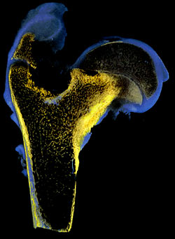
In 1977 Ruddick described the use of an ultraviolet fluorescence technique for improved tone separations in sections of bone. He cited Drury and Bullough in 1970 but took their technique a little further in order to demonstrate the difference between bone, soft tissue, and polyethylene and acrylic specimens. In particular he noted that it was possible to see where bone was extended into acrylic cement; this was used as part of a three-year research project on the study of implantation of prostheses. In 1979 he did a short review article for the fluorescence method - apart from the usual range of applications he also discussed the use of ultraviolet fluorescence in research to Perthes' disease (Figure 36).
References:
|
| © 2002 Prof. Robin Williams and Gigi Williams - Disclaimer URL: http://www.medicalphotography.com.au/Article_04/ Last modified: 3 May 2002 |