about | articles | authors | contact | links
about | articles | authors | contact | links |
![]() Home > Articles > Pioneers of invisible radiation photography > Lou Gibson
Home > Articles > Pioneers of invisible radiation photography > Lou Gibson
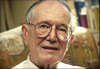
PIONEERS OF INVISIBLE RADIATION PHOTOGRAPHYAuthors: Prof. Robin Williams and Gigi Williams Lou Gibson (1906 - 1992)H. Lou Gibson (Figure 20) was born in Truro, Cornwall in 1906 and died in Rochester, New York in 1992. He was for many years, editor and consultant in medical, biological, scientific, and technical photography for the Eastman Kodak Company, received his B.Sc. degree in physics from the University of Illinois. He was contributing member and a fellow of the Biological Photographic Association, as well as a registered biological photographer of the BPA in the medicine specialty. He served as president, vice-president, and editor of the BPA Journal. In 1964 he won the Communications Award of the BPA for original work in infrared colour photography and medicine. Gibson's other photographic honors included the Combined Royal College's Medal of the Royal Photographic Society, the Louis B. Schmidt Award of the BPA, and the 1973 Progress Award and the Maxwell Trophy from the Photographic Society of America.
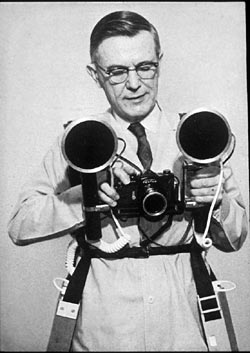
Figure 20 (above). Lou Gibson. Gibson (1945) wrote a general review paper on infrared photography of patients that included a helpful description of a practical technique and an evaluation of the clinical usefulness. Gibson described the use of photo-flash (Figure 21), the need to test camera equipment for infrared leakage, the requirement for even 'wrap-around' lighting, and the technique of unsharp area masking for accentuating fine detail. Within this paper was a comment by SM Bouton who presented the first application of infrared to the detection of cancer in the female breast. The photographs, which were not published till 1945, showed an asymmetrical pattern in cancerous breasts.
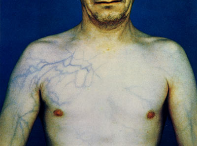
In 1961, Gibson published an interesting application of infrared in a paper entitled 'postprandial intensification of venous pattern' in Medical Radiography and Photography. In this presentation he demonstrated how the superficial venous pattern in a 12-year-old girl became greatly intensified following lunch. He suggested caution in interpreting venous patterns of the trunk and abdomen in view of this abnormal finding (Figure 22). Gibson presented this intriguing anomaly in the hope that other workers would attempt to use infrared to investigate other abnormalities in the vascular system.
Figure 22 (above). Intensification of superficial venous pattern following lunch. In a series of papers in the journal Medical and Biological Illustration, extending throughout 1962 and 1963, Lou Gibson described in great detail the technique of infrared luminescence, that is illuminating the subject with visible radiation and then recording the luminescence in the infrared part of the spectrum. Gibson presented extensive details of the technique and then its application to a wide range of medical and biological subjects. He presented a helpful table of a wide range of substances and it is interesting to note that bilirubin, for example, demonstrated a very strong infrared fluorescence, whereas biliverdin demonstrated none. He also claimed that it is easy to differentiate between jaundice and Addison's disease and Addison's disease in carotenaemia. Little work has been done since these pioneering publications. This may be due to the difficult nature of applying these techniques to the living subject. Continuing his work on diffuse lighting for infrared, in 1964 Gibson described the use of a white cubicle for achieving diffuse illumination of patients. He recommended the routine application of the technique as it provided a clearer 'map' of the veins and worked for any body region. Gibson presented another key publication in combination with two other researchers Buckley and Whitmore in 1965. This paper entitled 'new vistas in infrared photography for biological surveys' was published in the Journal of the Biological Photography Association. There were many points in this article worth noting, as always Gibson was assiduous in his research and presented all the references of previous work. He even challenged the, by then, sacrosanct view that arterial blood reflected infrared and presents evidence to show that it may not be the blood itself but the walls of the vessel that reflect the infrared. He photographed mesenteric veins and arteries exposed in a nitrogen atmosphere and found that the venous blood actually recorded lighter than the arterial blood. He also suggested the use of infrared Ektachrome film for the identification of haemosiderin. He demonstrated that melanin could be usefully distinguished from venous blood by the use of the infrared Ektachrome technique. Again he presented a very comprehensive table of different tissues and the colours that they reproduce using the infrared Ektachrome technique (Figure 23). He also claimed that there was a difference between oxyhaemoglobin and the reduced haemoglobin in their colour rendition in the infrared Ektachrome and suggested that this may be very helpful in looking at circulatory patterns. He presented many photographic examples. In an appendix to his paper, almost as an afterthought, Gibson described an experimental set-up in their department in which they constructed a 7ft tent in which the patient stood in order to achieve diffuse illumination for infrared photography - this was a development of his earlier idea here he used white hospital sheets to reflect off the light sources.
Figure 23 (above). Colour reproduction of infrared Ektachrome film. In 1967, Gibson published a two-part work in the journal Visual Medicine; this was entitled 'medical infrared colour photography'. Whilst it contained many of the techniques and applications published in his early career, the second part entitled 'applications' contained some exciting new observations. For instance he hinted that the use of infrared for the recording of malignant neoplasms might be useful not because of the absorbency of infrared by the pigment, but because of the increased absorption due to the greater blood supply. He also challenged the accepted view that infrared radiation penetrated to a depth of 3mm. He stated clearly that he could find no account of experiments to test what might be called 'the photographic penetration of infrared radiation'. Almost all previous workers having tested skin in vitro with spectrophotometers. The author described research undertaken where he inserted carbon rods of various thicknesses underneath the prepuce; he was able to demonstrate these with infrared photography. References:
|
| © 2002 Prof. Robin Williams and Gigi Williams - Disclaimer URL: http://www.medicalphotography.com.au/Article_04/ Last modified: 3 May 2002 |