about | articles | authors | contact | links
about | articles | authors | contact | links |
![]() Home > Articles > Pioneers of invisible radiation photography > Leo Massopust
Home > Articles > Pioneers of invisible radiation photography > Leo Massopust
PIONEERS OF INVISIBLE RADIATION PHOTOGRAPHYAuthors: Prof. Robin Williams and Gigi Williams Leo Massopust (1893 - 1970)Leo Massopust (Figure 18) was a biological photographer, an accomplished landscape artist and musician. He spent his working life at Marquette University School of Medicine as medical artist and photographer. He served for almost twenty years as the Editor of the Journal of Biological Photography and was past President of the Biological Photographic Association. He described his experimental infrared technique in two papers in 1934, one in Radiography and Clinical Photography and the other in the Anatomical Record.
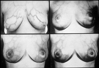
In 1936 Massopust published a paper on 'Infrared photographic study of the changing pattern of the superficial veins in the case of human pregnancy.' This publication in the journal Surgery, Gynecology and Obstetrics marked the beginning of the application of infrared photography to the female breast, indeed the author described in his second paragraph the technique he claimed to have had in regular use since 1933. From his infrared photography, Massopust concluded that there was an increase in the size and conspicuousness of the superficial vessels in the breast and abdomen during pregnancy and claimed it could be used as a pregnancy test. The story of infrared recording of the breast is complex. It is the authors' belief that Massopust was the first to apply the technique to looking at superficial veins of the female breast but that the first application of infrared recording to the early detection of cancer of the breast was actually by Gibson in 1944. Whilst working at the St Mary's Hospital Rochester, Gibson was able to detect asymmetrical patterns in the breasts of women who turned out to have breast tumours. His findings were not reported until 1945. Massopust published another paper in 1937 entitled 'infrared photography of gross anatomical specimens'. He used the technique reported three years earlier on specimens of the lung and human placenta, he discussed the techniques of injecting the specimens with red cinnabar and black India ink to get better contrast in the infrared results. He concluded by saying that infrared photography was of great value in revealing the morphological detail of gross anatomical specimens. Massopust (1945) published a tutorial article on infrared photography including recommendations for films, filters, processing and lighting. He gave various hints about the necessity for checking for example, bellows and plate holders and advised focusing with a Wratten 25 red filter in order to reduce the effects of focus shift. In 1948, in the journal Surgery, Gynecology and Obstetrics, Massopust presented his first findings in relation to the superficial veins and early detection of breast tumours. This preliminary report included some 100 cases. Two years later in 1950 in the same journal, Massopust and Gardner presented the results of over 1000 cases for consideration (Figure 19). In this paper they suggested an anatomical classification for the superficial venous patterns of the thorax and made some observations on the principal drainage of the mammary glands. They stated that further studies were needed before any exact clinical correlation could be made between the venous pattern and the state of the breast. Three years on Massopust and Gardner had collected 1200 cases and in their last paper in the series in the same journal they then made some observations about the value of infrared photography in detecting breast cancer. Massopust was lukewarm in his recommendation that infrared photography could act 'as an additional aid to the physician in the differentiation of breast tumours.' In this study of 1200 cases there were 12 where the infrared phlebogram disagreed with the final clinical diagnosis, but 381 incidences where an accurate clinical diagnosis was made without surgical intervention.
Figure 19 (above). The use of infrared photography for the early detection of breast tumors. References:
|
| © 2002 Prof. Robin Williams and Gigi Williams - Disclaimer URL: http://www.medicalphotography.com.au/Article_04/ Last modified: 3 May 2002 |