about | articles | authors | contact | links
about | articles | authors | contact | links |
![]() Home > Articles > Reflected Ultraviolet Photography > Electronic recording of the ultraviolet image
Home > Articles > Reflected Ultraviolet Photography > Electronic recording of the ultraviolet image
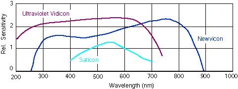
REFLECTED ULTRAVIOLET PHOTOGRAPHYAuthors: Prof. Robin Williams and Gigi Williams Electronic recording of the ultraviolet imageWhile the photographic recording of the reflected ultraviolet image undoubtedly yields higher quality, there is a role for electronic visualization of the subject in real time. The eye is not sensitive to ultraviolet reflected from the subject and often we do not know whether there is anything of interest to photograph. It can be helpful, therefore, to have some form of video pre-visualization before proceeding to full photographic recording. Mustakallio and Korhonen (1966) for example, used an ultraviolet sensitive television tube to examine dermatological conditions, as did Marshall in 1981. Anselmo and Zawaki (1973) used a multi-channel television system in the assessment of burns, and Morton and Miller (1981) used the infrared sensitivity of a Newvicon tube to assess breast lesions prior to photography. Conventional video tubes are not sensitive to the ultraviolet region but there are specialized tubes, the GE 8507 for example, which are sensitive down as far as 150nm (these often have quartz faceplates and must be used with quartz and silica lenses). Figure 56 shows the spectral sensitivity of Newvicon and Ultraviolet Silicon Vidicon tubes compared to the normal Saticon tube commonly found in analogue television cameras. Most manufacturers supply ultraviolet sensitive tubes, for example GE's 8507 and Z79125, RCA's C23231 and 4532, and EMI's 9677UV.
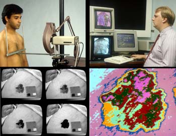
Figure 56 (above). The spectral sensitivity of Newvicon and Ultraviolet Silicon Vidicon tubes compared to the normal Saticon tube found in analogue television cameras. Where the reflected image is of very low brightness it is possible to use an ultraviolet image intensifier tube. The Hamamatsu V3229P, for example, is sensitive within the range 200 to 320nm and enables the very weakest of images to be recorded. Solid state charge coupled devices (CCDs) are also now available with resolution and sensitivity in the ultraviolet region to match that of silicon target tubes. The COHU Corporation 4800 series, for example, have a peak sensitivity at 285nm. Murray (1988) used the output from an ultraviolet sensitive CCD camera in the assessment of change in pigmented lesions of the skin. He digitized the television image and then used customized image analysis software to measure changes in the shape and colour of the lesions. (Figure 57). Analysis of the digital image extends the usefulness of invisible radiation techniques.
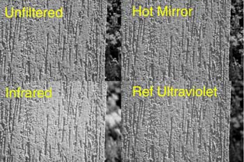
Figure 57 (above). Murray's early (1988) CCD camera system. He digitized the television image and then used customized image analysis software to measure changes in lesions. The widespread use of digital cameras, and the popular discovery that the CCDs in these cameras are sensitive to both ultraviolet and infrared, has caused a resurgence of interest in photography with invisible radiation: although it has to be said that most are 'novelty' pursuit rather than robust professional solutions. All the CCDs used in consumer digital cameras are sensitive to ultraviolet. Unfortunately the CCDs are also sensitive to infrared and the standard ultraviolet transmission filter (Wratten, Schott or B+W) has a significant window in the infrared - a fact overlooked by most practitioners. The recording of 'pure' reflected ultraviolet with a digital camera therefore requires the addition of an infrared absorbing filter in addition to the "Wood's" filter. These infrared absorption filters are often called "hot filters." There is some question as to whether the auto-focus facility of digital cameras is accurate outside of the visible spectrum. The authors tested a Nikon 990 with infrared absorbing, infrared transmission and ultraviolet transmission filters and found that the autofocus facility worked satisfactorily (Figure 58); but as with all these issues it is probably best to test your own digital camera in this same way. If the camera's autofocus will not function - as a result of either the wavelength shift or the low illumination levels - it may still be possible to switch to manual focussing. Certainly, where the digital's autofocus capability is accurate this represents a very significant advantage for electronic imaging over film based recording.
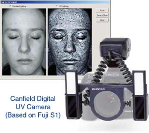
Figure 58 (above). The autofocus mode of the Nikon 990 digital camera evaluated. The images of a rendered wall demonstrate that the Nikon copes with each wavelength selected. (NB The ultraviolet record was imaged by combining an infrared absorbing filter - 'hot filter' - with the Wratten 18A. When the ultraviolet filter alone is used the camera is compromising between the accurate focus for ultraviolet and that for infrared.) The sensitivity of different CCDs varies enormously and it is impossible therefore to give any useful recommendations for exposure etc., except to say that exposures are likely to be very long - often several seconds with most continuous sources of radiation. This causes two problems: firstly, the obvious problem of camera and/or subject movement and secondly, noise from the CCD in low light conditions. The solution to movement is electronic flash - preferably without any UV coating (see sources of ultraviolet radiation) or a tripod for subjects that will not move. The second problem - noise- is caused by an aberration of the charged couple device. The electronics themselves generate heat and this energy causes some pixel electrons to be activated and then trapped as 'false positives' ie. not generated by light energy falling on the pixel. This results in a blotchy image in dark shadow areas of the image: it becomes worse in hot conditions and with very long exposures. CCDs built into astronomical imaging devices (which have very long exposure times) are often super cooled in order to avoid this problem of noise. It is possible to 'filter' out this noise at a post imaging stage in an image manipulation software program such as Adobe Photoshop by a technique known widely in astronomy as dark field subtraction. Two images are required - one of the reflected ultraviolet record (with noise) and one with no ambient illumination (the noise). The image of the noise is then effectively subtracted from the real image using Photoshop's layer subtraction feature. Canfield Scientific in the US supply a complete digital UV kit based on the Fuji S1 digital camera (Figure 59). This system features dual flashes and an integral filter system - it is designed for ease of use in the clinic or surgery on subjects up to approximately 1:10 reproduction ratio (the low power of the flash preventing adequate illumination of larger subjects).
Figure 59 (above). Canfield Scientific in the US supply a complete digital UV kit based on the Fuji S1 digital camera. References
|
| © 2002 Prof. Robin Williams and Gigi Williams - Disclaimer URL: http://www.medicalphotography.com.au/Article_01/ Last modified: 3 May 2002 |