about | articles | authors | contact | links
about | articles | authors | contact | links |
![]() Home > Articles > Pioneers of invisible radiation photography > Ray Lunnon
Home > Articles > Pioneers of invisible radiation photography > Ray Lunnon
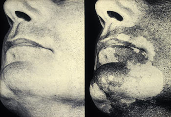
PIONEERS OF INVISIBLE RADIATION PHOTOGRAPHYAuthors: Prof. Robin Williams and Gigi Williams Ray LunnonA significant step forward in the use of ultraviolet radiation in biomedical photography was made in 1959 when the use of electronic flash was first described. Two authors, working at the same time but quite independently, described how electronic flash could be used for reflected ultraviolet photography. Birgitta Lindenstam described how it was useful for photographing teeth whilst Ray Lunnon reported the first use of electronic flash for recording skin. He showed examples of both depigmentation in vitiligo (Figure 26) and hyperpigmentation in a pigmented naevus and noted the fine surface detail of skin. In his discussion he pointed out the significant advantages of the use of electronic flash, and also stressed the importance of always including a visible light photograph alongside any photograph taken in invisible radiation. This advice still holds good to date. He also noted that the negatives produced by the direct ultraviolet photographic technique had a lower contrast ratio and needed an increase in development by 30% to achieve sufficient contrast.
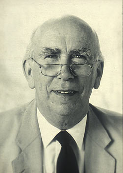
Figure 26 (above). Ultraviolet photograph showing depigmentation in vitiligo. Lunnon (Figure 27) went on to become one of the most accurate and consistent authors in the field of direct reflected ultraviolet photography, and has a series of publications (Lunnon 1961, 1968, 1976, 1979) showing various conditions of the skin and eyes. In his 1961 paper on the photography of diseases of the skin and again in 1968 Lunnon showed an example of tinea capitas fluorescing under ultraviolet radiation. Also in the 1961 paper he briefly mentioned infrared photography and showed an example of how venous blood absorbed infrared making this technique useful for venous engorgement, or collateral circulation. In 1968 he showed examples of pooling of fluorescein dye under ill-fitting contact lenses.
Lunnon was the first person in the United Kingdom to obtain a higher degree in medical photography and his work was based on his reflected ultraviolet techniques. The Faculty of Medicine of the University of London accepted his thesis entitled 'reflective ultraviolet photography in medicine', in May 1974 for the degree of Master of Philosophy. Lunnon tested everything by original experimentation and revealed several new observations. In particular he noted that the focus shift in ultraviolet was quite contrary to that which had been quoted by previous authors, and that it went in the same direction as infrared when one is considering complex compound lenses. He was probably the first author to do an authoritative and complete literature review and revealed a number of historical sources ignored by other workers. In one chapter of the thesis alone, he described over 120 original experiments depicting the application of reflected ultraviolet technique to clinical conditions. He showed examples of hyperpigmentation that were more clearly delineated in the reflected ultraviolet technique such as freckles, Peutz-Jegher syndrome, Berlocque dermatitis, melasma and naevus pigmentosus. He showed examples of hypopigmentation such as vitiligo, alopecia areata of beard, Sutton's halo naevus, scleroderma, psudeoxanthoma-elasticum, tuberous sclerosis and adenoma sebaceum. In addition he showed examples where the surface detail was more defined and where nails and vascular conditions could be shown better such as in lichenoid eczema, palmar skin ridging, contact dermatitis, nummular eczema, molluscum contagiosum, nail dystrophy, scleroderma and trauma to the hand, erythema and livedo. He also showed many examples of eyes. He found that most irises had the same reflectivity in the ultraviolet record but that the muscle structure could be revealed, as could any melanomatas. He showed that the ultraviolet record could show greater detail in the albino iris and that in the normal eye very high definition of scleral and conjuctival vessels could be seen. References:
|
| © 2002 Prof. Robin Williams and Gigi Williams - Disclaimer URL: http://www.medicalphotography.com.au/Article_04/ Last modified: 3 May 2002 |