
about | articles | authors | contact | links
about | articles | authors | contact | links |
![]() Home > Articles > Infrared Photography > Biomedical applications gallery
Home > Articles > Infrared Photography > Biomedical applications gallery
INFRARED PHOTOGRAPHYAuthors: Prof. Robin Williams and Gigi Williams Biomedical applications of infrared photography:
|
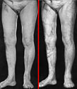 Figure 58. Varicose veins. |
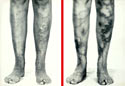 Figure 59. Sub-cutaneous veins. |
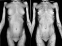 Figure 60. Postprandial engorgement of the veins. |
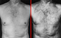 Figure 61. Collateral circulation around mediastinal tumour. |
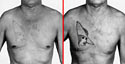 Figure 62. Another example of collateral circulation. |
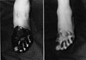 Figure 63. Penetration of burn escha. |
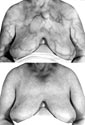 Figure 64. Venous blood return from breasts. |
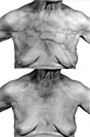 Figure 65. Uneven vascular patterns in neoplastic disease. |
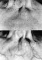 Figure 66. Vascular patterns around a pulsating tumour. |
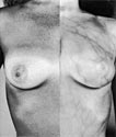 Figure 67. Subcutaneous veins. |
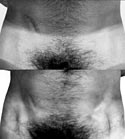 Figure 68. Transparency of cutaneous melanin. |
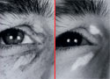 Figure 69. Transparency of melanin in the ephiledes. |
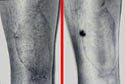 Figure 70. Melanin in malignant melanoma. |
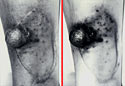 Figure 71. Same, three months later. |
|
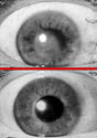 Figure 72. Penetration of lens opacity. |
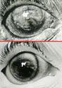 Figure 73. Penetration of severe corneal opacity. |
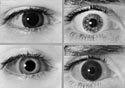 Figure 74. Dark brown compared to blue irises. |
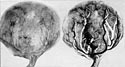 Figure 75. Human placenta injected with red cinnabar into arteries and black india ink into veins |
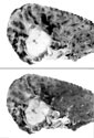 Figure 76. Silicotic deposits in the lung. |
|
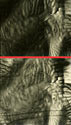 Figure 77. IR visibility of burned document. |
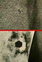 Figure 78. Firearm powder burns on fabric. |
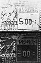 Figure 79. Forgery of a postal meter stamp. |
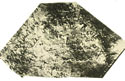 Figure 80. Pressure imprint of writing. |
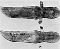 Figure 81. Knife sheath with hidden detail. |
 Figure 82. Text obliterated by dye type ink. |
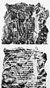 Figure 83. Dead Sea Scrolls. |
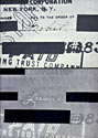 Figure 84. IR examination of a document. |
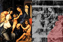 Figure 85. IR examination of a painting. |
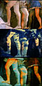 Figure 86. Restoration of a painting. |
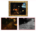 Figure 87. Detection of artist's signature. |
|
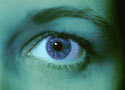 Figure 88. Melanin with Infrared Ektachrome film. |
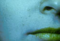 Figure 89. Lipstick and skin with Infrared Ektachrome. |
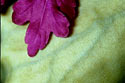 Figure 90. Chlorophyll with Infrared Ektachrome. |
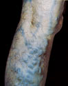 Figure 91. Deoxygenated blood. |
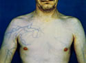 Figure 92. Collateral circulation. |
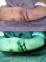 Figure 93. Bruises examined at post mortem. |
| © 2002 Prof. Robin Williams and Gigi Williams - Disclaimer URL: http://www.medicalphotography.com.au/Article_03/ Last modified: 3 May 2002 |