about | articles | authors | contact | links
about | articles | authors | contact | links |
![]() Home > Articles > Infrared Photography > Reflected infrared photography: The practical operating "window"
Home > Articles > Infrared Photography > Reflected infrared photography: The practical operating "window"
INFRARED PHOTOGRAPHYAuthors: Prof. Robin Williams and Gigi Williams Reflected infrared photography
|
||||||||||||||||||||||||||||||||||||||||||||||||||||||||||||||||||||||||||||||||||
| Method | Black & White Infrared | Control | ||
| Film | Kodak B&W HIE 135 | T-Max 100 or other | ||
| Processing | ID-11 stock - 11 mins @20°C | T-Max Developer - 8 mins 20°C | ||
| Filter | 88A | None | ||
| Lens | Old 105mm Micro Nikkor | 105mm Micro Nikkor | ||
| Focus Shift | Move the whole camera back by: 1:10 53mm 1:8 32mm 1:4 8mm 1:2 5mm 1:1 2mm |
None | ||
| Light Source | Studio flash with light form panels | SB-140 | Studio flash with light form panels | SB-140 |
| Exposures | Meter for 400 ISO | 1:10 f16 Full Power 1:8 f11 1/4 Power 1:4 f11/16 1/4 Power 1:2 f16 1/4 Power 1:1 f16 1/4 Power |
Meter as usual | Full Power 1:10 f11 1:8 f11/16 1:4 f16 1:2 f22 1:1 f22 |
| Method | Colour Infrared | Control | ||
| Film | Kodak EIR | Ektachrome 100 or other | ||
| Processing | E-6 | E-6 or other | ||
| Filter | Wratten 12 | None | ||
| Lens | 105mm Micro Nikkor | 105mm Micro Nikkor | ||
| Focus Shift | None | Not necessary | ||
| Light Source | Studio flash | SB-140 | Studio flash | SB-140 |
| Exposures | Meter for 200 ISO if using Wratten 12 | 1:10 f16 Full Power 1:8 f11 Full Power 1:4 f11/16 Full Power 1:2 f16 Full Power 1:1 f11 Full Power |
Meter as usual | Full Power 1:10 f11 1:8 f11/16 1:4 f16 1:2 f22 1:1 f22 |
It is critically important that a standard 'control' image - in colour or panchromatic black-and-white - that represents the visual appearance of the subject, be included as a reference with all scientific invisible radiation records. Obtaining exactly the same viewpoint, with the same lighting can be a very challenging task; this is exacerbated still further when undertaking multi-spectral analysis, where at least three cameras are involved. The authors designed a simple system (Figure 50) where the electronic flash is actually fixed to a tripod and quick release plates fitted to tripod and each camera allow the rapid change over of cameras.
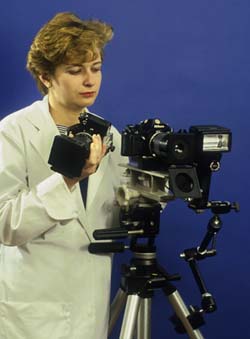 |
Figure 50 (left). A practical working arrangement for multispectral analysis. The flash is mounted to the tripod and remains in a constant position whilst cameras are changed easily by utilizing quick release plates. |
| © 2002 Prof. Robin Williams and Gigi Williams - Disclaimer URL: http://www.medicalphotography.com.au/Article_03/ Last modified: 3 May 2002 |