
about | articles | authors | contact | links
about | articles | authors | contact | links |
![]() Home > Articles > Reflected Ultraviolet Photography > Biomedical applications gallery
Home > Articles > Reflected Ultraviolet Photography > Biomedical applications gallery
REFLECTED ULTRAVIOLET PHOTOGRAPHYAuthors: Prof. Robin Williams and Gigi Williams Biomedical applications of reflected ultraviolet photography:
|
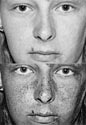 Figure 63. Delineation of ephelides. |
 Figure 64. Detailed view of ephelides. |
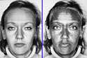 Figure 65. Increase in melanin. |
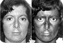 Figure 66. An example of melasma. |
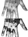 Figure 67. Reduction of melanin in vitiligo. |
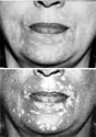 Figure 68. Less obvious case of vitiligo. |
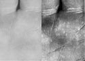 Figure 69. Surface detail of skin. |
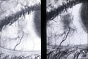 Figure 70. Surface detail in episcleritis. |
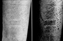 Figure 71. Delineation of contact dermatitis. |
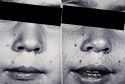 Figure 72. A case of Molluscum contagiosum. |
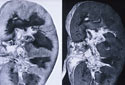 Figure 73. Specimen photography. |
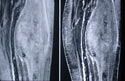 Figure 74. Specimen of neuroblastoma. |
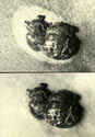 Figure 75. Sutton's pigmented lesion of the skin. |
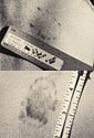 Figure 76. Detection of bruising. |
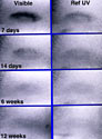 Figure 77. A bruise over three months. |
 Figure 78. Detection of forgery. |
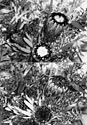 Figure 79. Proteas. |
Figure 80. A selection of flowers. |
< Biomedical applications for reflected ultraviolet photography |
| © 2002 Prof. Robin Williams and Gigi Williams - Disclaimer URL: http://www.medicalphotography.com.au/Article_01/ Last modified: 3 May 2002 |